39 light microscope with labels
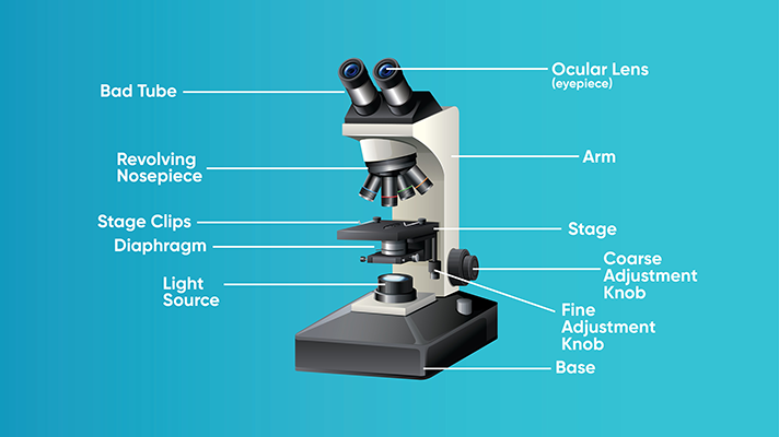
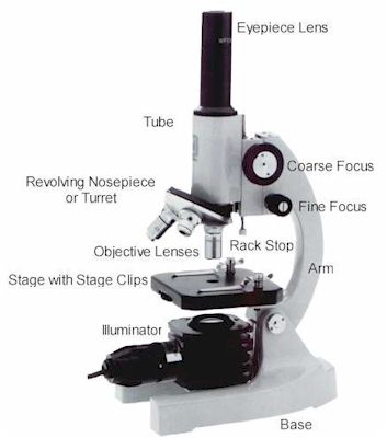
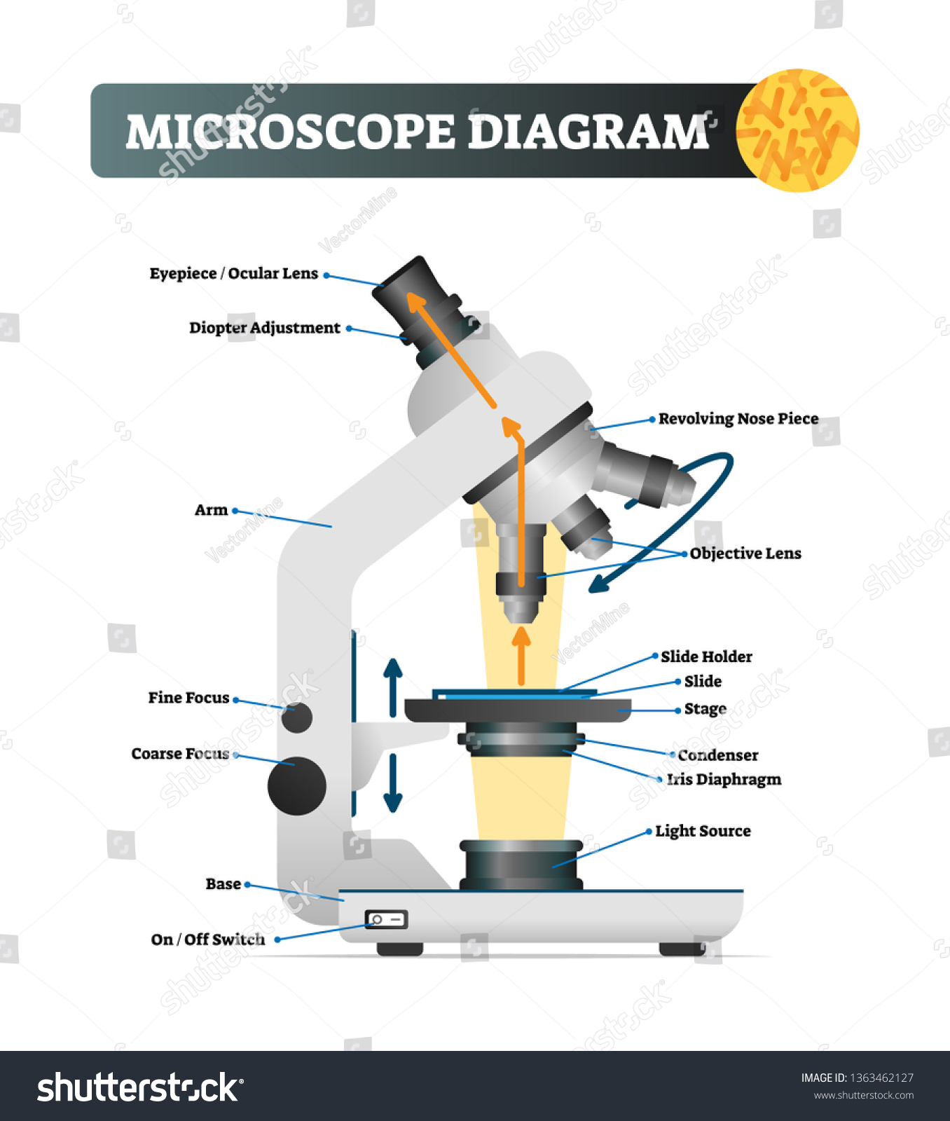
Simple Microscope - Diagram (Parts labelled), Principle, Formula and Uses The magnification power of a simple microscope is expressed as: M = 1 + D/F Where M = Magnification power D = the lease possible distance of distinct vision of eye, typically 25cm F = Focal length of the convex lens It is to be noted that Label the Light Microscope - Labelled diagram Eyepiece, Light Source, Base, Stage, Stage Clips, Fine Focus, Coarse Focus, Arm, Objective Lens, Diaphragm.
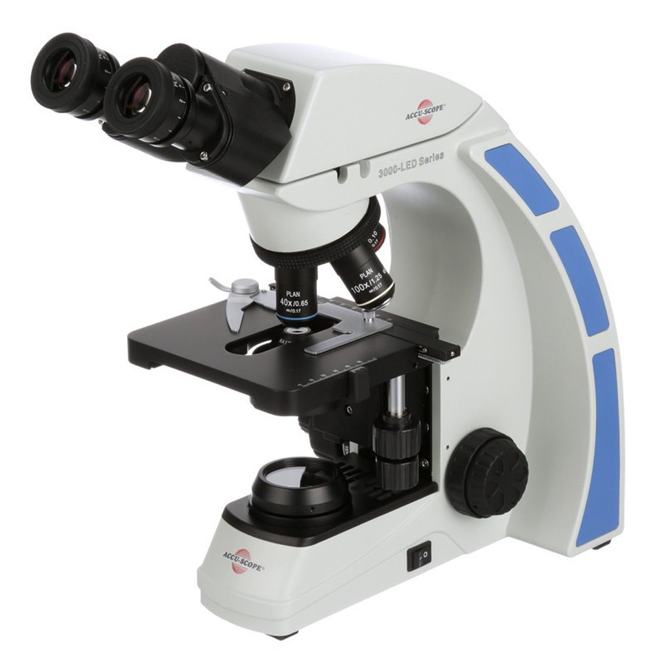
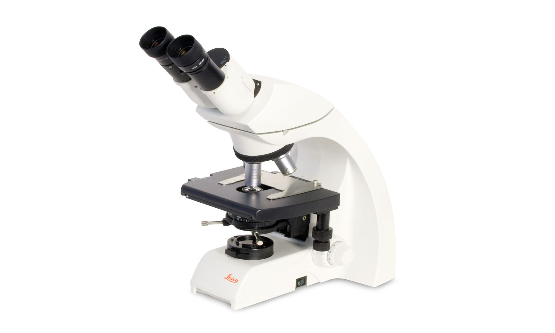
Compound Light Microscope: Everything You Need to Know A compound light microscope is a type of light microscope that uses a compound lens system, meaning, it operates through two sets of lenses to magnify the image of a specimen. It's an upright microscope that produces a two-dimensional image and has a higher magnification than a stereoscopic microscope.

Light microscope with labels
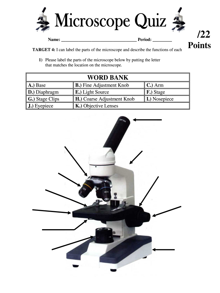
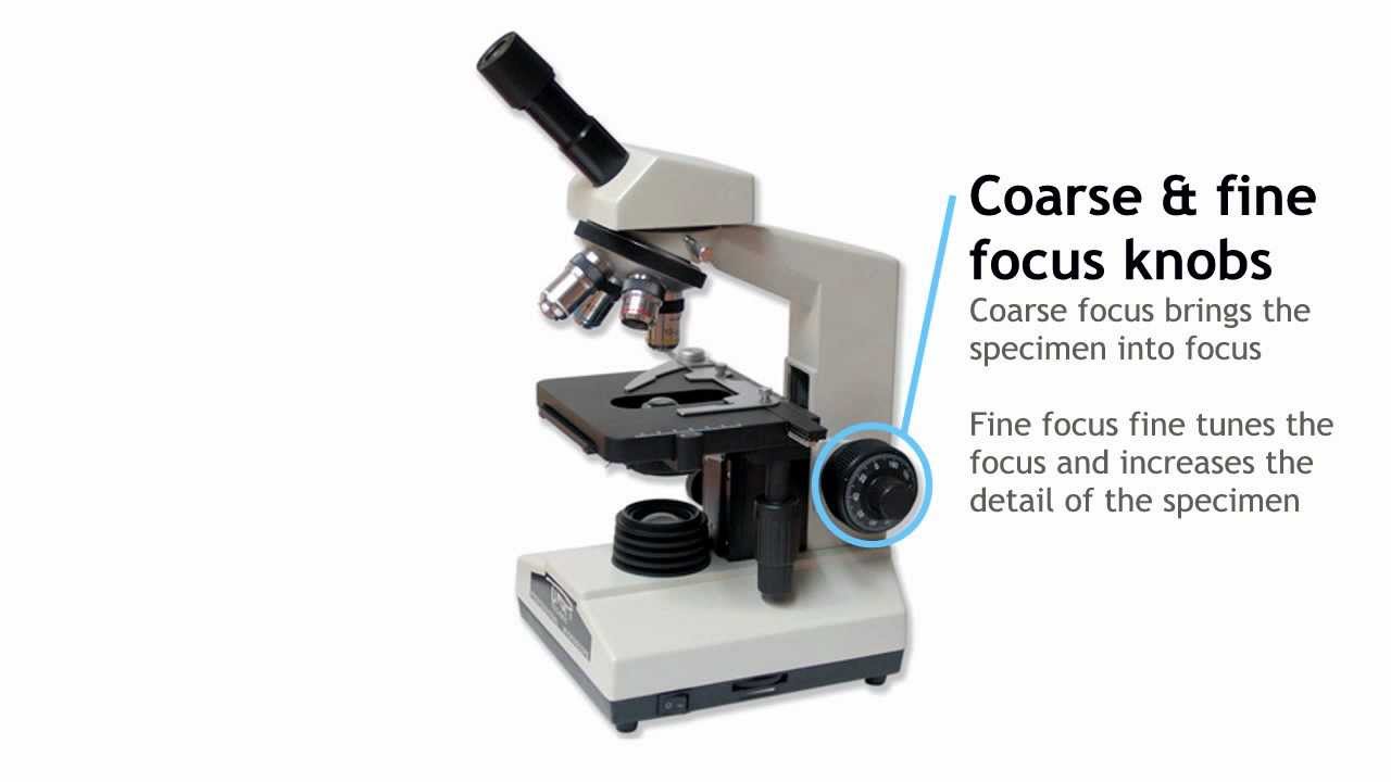
Labeling the Parts of the Microscope | Microscope World Resources Labeling the Parts of the Microscope This activity has been designed for use in homes and schools. Each microscope layout (both blank and the version with answers) are available as PDF downloads. You can view a more in-depth review of each part of the microscope here. Download the Label the Parts of the Microscope PDF printable version here. Compound Microscope Parts, Functions, and Labeled Diagram Compound Microscope Definitions for Labels Eyepiece (ocular lens) with or without Pointer: The part that is looked through at the top of the compound microscope. Eyepieces typically have a magnification between 5x & 30x. Monocular or Binocular Head: Structural support that holds & connects the eyepieces to the objective lenses. Light Microscope: Functions, Parts and How to Use It To use a light microscope, you can follow the steps below carefully. Start with a low lens and a clean slide. The microscope stage should be lowered as low as possible. Center the slide so that the specimen is under the objective lens. Use the coarse adjustment knob to get a general focus. Then slowly move up the stage until focus is achieved.
Light microscope with labels. Microscope Parts and Functions The specimen is placed on the glass and a cover slip is placed over the specimen. This allows the slide to be easily inserted or removed from the microscope. It also allows the specimen to be labeled, transported, and stored without damage. Stage: The flat platform where the slide is placed. Microscope Labeling Game - PurposeGames.com About this Quiz. This is an online quiz called Microscope Labeling Game. There is a printable worksheet available for download here so you can take the quiz with pen and paper. This quiz has tags. Click on the tags below to find other quizzes on the same subject. Science. City of Calgary (@cityofcalgary) / Twitter Aug 21, 2008 · Official City of Calgary local government Twitter account. Keep up with City news, services, programs, events and more. Not monitored 24/7. Label the Light Microscope - Labelled diagram - Wordwall Drag and drop the pins to their correct place on the image.. Eyepiece, Light Source, Base, Stage, Stage Clips, Fine Focus, Coarse Focus, Arm, Objective Lens.
A Study of the Microscope and its Functions With a Labeled Diagram ... Light Microscopes: These use light rays to illuminate objects. e.g. Dissection microscopes and compound microscopes. Electron Microscopes: These illuminate objects with a beam of highly charged electrons. e.g. Transmission electron microscope (TEM) and scanning electron microscope (SEM). Compound Microscope - Diagram (Parts labelled), Principle and Uses See: Labeled Diagram showing differences between compound and simple microscope parts Structural Components The three structural components include 1. Head This is the upper part of the microscope that houses the optical parts 2. Arm This part connects the head with the base and provides stability to the microscope. Light Microscope- Definition, Principle, Types, Parts, Labeled Diagram ... Brightfield Light Microscope (Compound light microscope) This is the most basic optical Microscope used in microbiology laboratories which produces a dark image against a bright background. Made up of two lenses, it is widely used to view plant and animal cell organelles including some parasites such as Paramecium after staining with basic stains. Microscope Labeling - The Biology Corner Microscope Labeling. Shannan Muskopf May 31, 2018. This simple worksheet pairs with a lesson on the light microscope, where beginning biology students learn the parts of the light microscope and the steps needed to focus a slide under high power. The labeling worksheet could be used as a quiz or as part of direct instruction where students label the microscope as you go over what each part is used for.
PDF Parts of the Light Microscope - Science Spot Supports the MICROSCOPE D. STAGE CLIPS HOLD the slide in place C. OBJECTIVE LENSES Magnification ranges from 10 X to 40 X F. LIGHT SOURCE Projects light UPWARDS through the diaphragm, the SPECIMEN, and the LENSES H. DIAPHRAGM Regulates the amount of LIGHT on the specimen E. STAGE Supports the SLIDE being viewed K. ARM Used to SUPPORT the Could Call of Duty doom the Activision Blizzard deal? - Protocol Oct 14, 2022 · “The CMA is concerned that having full control over this powerful catalogue, especially in light of Microsoft’s already strong position in gaming consoles, operating systems, and cloud infrastructure, could result in Microsoft harming consumers by impairing Sony’s — Microsoft's closest gaming rival — ability to compete,” the report ... Light Microscope Parts, Function & Uses - Study.com The following are the most common components found on light microscopes: Microscope parts Ocular lenses: Allow the viewer to look into the microscope, usually 10x magnification Revolving... Compound Light Microscope Labelling Quiz - PurposeGames.com This is an online quiz called Compound Light Microscope Labelling. There is a printable worksheet available for download here so you can take the quiz with pen and paper. Your Skills & Rank. Total Points. 0. Get started! Today's Rank--0. Today 's Points. One of us! Game Points. 15.
Microscope Maintenance Tips | Science supplies, Microscope ...
Parts of the Microscope with Labeling (also Free Printouts) Parts of the Microscope with Labeling (also Free Printouts) By Editorial Team March 7, 2022 A microscope is one of the invaluable tools in the laboratory setting. It is used to observe things that cannot be seen by the naked eye. Table of Contents 1. Eyepiece 2. Body tube/Head 3. Turret/Nose piece 4. Objective lenses 5. Knobs (fine and coarse) 6.

National 131-RLED-MS Compound Microscope with Mechanical Stage
ZEISS Axio Observer for Life Science Research Fast switchable light sources and filters give you highest spectral flexibility and speed. Select the ideal camera to always get the image quality and speed your applications require. Whether keeping your sample in focus for long-term imaging or adapting your objective to your sample, it's all automatic with this highly organized system.

2.2 Molecular make up of cells | Cells: the basic units of ...
Binocular Microscope Anatomy - Parts and Functions with a Labeled ... Now, I will describe all these non-optical parts of the light compound microscope with the labeled diagrams. The body tube of the microscope. The body tube is the solid support for the optical and mechanical parts of the microscope. There are two basic types of stand in the body tube of a light compound microscope - upright stand and inverted ...

Microscope Diagram Labeled, Unlabeled and Blank | Parts of a ...
Electron microscope - Wikipedia An electron microscope is a microscope that uses a beam of accelerated electrons as a source of illumination. As the wavelength of an electron can be up to 100,000 times shorter than that of visible light photons, electron microscopes have a higher resolving power than light microscopes and can reveal the structure of smaller objects.
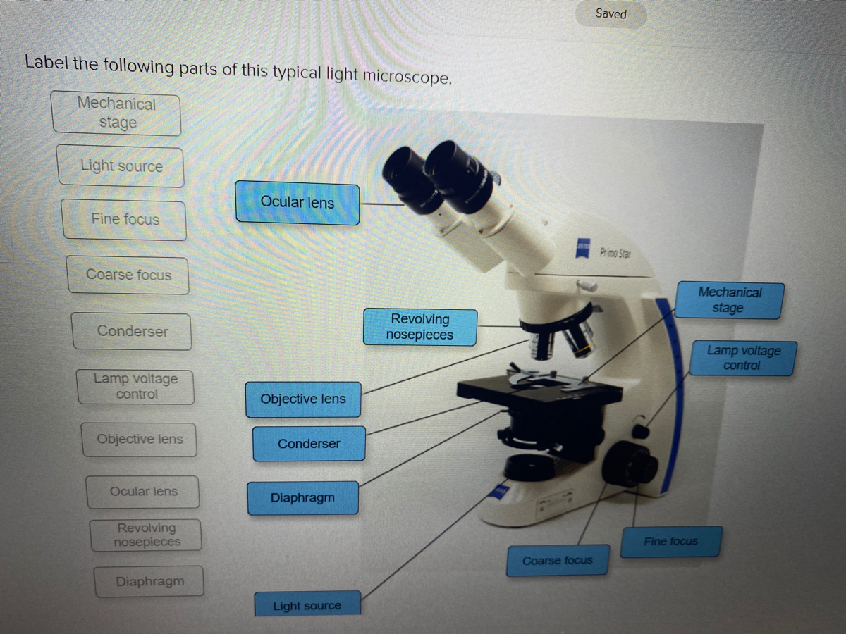
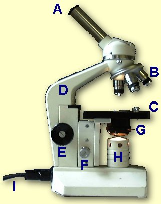
Answered: Saved Label the following parts of this… | bartleby
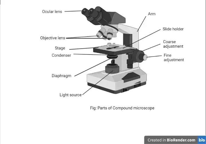
Compound Microscope Parts - Labeled Diagram and their Functions Always lift a microscope by holding both the arm and base with two hands. There are two major optical lens parts of a microscope: Eyepiece (10x) and Objective lenses (4x, 10x, 40x, 100x). Total magnification power is calculated by multiplying the magnification of the eyepiece and objective lens. The illuminator provides a source of light.

Light Microscope Worksheet worksheet
Microscope, Microscope Parts, Labeled Diagram, and Functions Revolving Nosepiece or Turret: Turret is the part of the microscope that holds two or multiple objective lenses and helps to rotate objective lenses and also helps to easily change power. Objective Lenses: Three are 3 or 4 objective lenses on a microscope. The objective lenses almost always consist of 4x, 10x, 40x and 100x powers. The most common eyepiece lens is 10x and when it coupled with ...
The Compound Light Microscope Label the following parts on ...
Shop by Category | eBay Shop by department, purchase cars, fashion apparel, collectibles, sporting goods, cameras, baby items, and everything else on eBay, the world's online marketplace
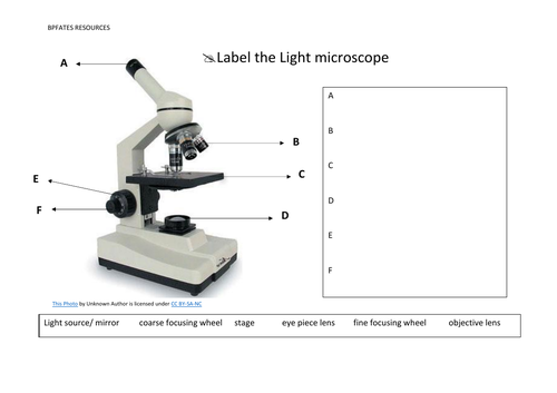
Label the light microscope | Teaching Resources
Lipid bilayer - Wikipedia Lipid bilayers cannot be seen in a traditional microscope because they are too thin. In order to see bilayers, researchers often use fluorescence microscopy. A sample is excited with one wavelength of light and observed in a different wavelength, so that only fluorescent molecules with a matching excitation and emission profile will be seen.

Parts of a Microscope with Their Functions – Microbe Online
Light microscopes - Cell structure - Edexcel - BBC Bitesize Microscopes are used to produce magnified images. There are two main types of microscope: light microscopes are used to study living cells and for regular use when relatively low magnification...

Parts of a microscope with functions and labeled diagram
Light Microscope Parts Labeled - 18 images - parts of the microscope ... [Light Microscope Parts Labeled] - 18 images - optical microscopy and specimen using the transmission, microscope with labels clip art at vector clip, solved microscope parts labeling 9 label the image of a c, ,

Labels for the light microscope for... - The Science Break ...
Amazon.com: AmScope 120X-1200X 52-pcs Kids Beginner ... Nov 18, 2013 · Buy AmScope 120X-1200X 52-pcs Kids Beginner Microscope STEM Kit with Metal Body Microscope, Plastic Slides, LED Light and Carrying Box (M30-ABS-KT51),Black: Microscopes - Amazon.com FREE DELIVERY possible on eligible purchases

microscope | Types, Parts, History, Diagram, & Facts | Britannica
Label the microscope — Science Learning Hub 8 Jun 2018 — Label the microscope · light source: Sends light onto the specimen/slide · fine focus adjustment: Moves the lens in order to make very small ...

Labeling the Parts of the Microscope | Microscope activity ...
Compound Microscope: Definition, Diagram, Parts, Uses, Working ... - BYJUS The compound microscope is also known as the bright-field microscope because the light passes directly through the light source to the eye through the two lenses. This mechanism makes the field of vision brightly illuminated. Parts of Compound Microscope

Ultimate Science Microscope Kit - My First Lab MFL-05 Cordless Compound Microscope for Students w/ 4X, 10x & 40X Eyepieces – Illuminated 40-400x ...
Parts of a Microscope - The Comprehensive Guide Step 1: Fully open field and condenser diaphragms and focus on specimen using x10 objective. Step 2: Fully close field diaphragm and adjust the condenser and focus so edges are as sharp as possible. Step 3: Use screws at front of condenser to centre field diaphragm and open field diaphragm to fill view. Step 4: Remove eyepiece and close down ...
Parts of a Microscope Quiz
Compound Microscope Labeled Diagram | Quizlet High-power objective lense (Magnification- the bigger one) contains the lenses with higher power of magnification Stage clips holds slides on stage Light source Projects light upwards through the diaphragm to allow you to see the specimen Diaphram Disk under the stage that allows light through. Base Bottom or lower part of the microscope Objectives
Parts of a Microscope with Their Functions – Microbe Online
Parts of a microscope with functions and labeled diagram - Microbe Notes Microscopic illuminator - This is the microscopes light source, located at the base. It is used instead of a mirror. It captures light from an external source of a low voltage of about 100v. Condenser - These are lenses that are used to collect and focus light from the illuminator into the specimen.

Parts of the Microscope with Labeling (also Free Printouts ...
Sperm Under Microscope with Labeled Diagram - AnatomyLearner Under the light microscope, the sperm consists of two main portions - the head and the tail. But, the electron microscope shows four different parts in the tail of spermatozoa. ... So, this article provides the details structural features of sperm under the light microscope. All the labeled diagrams might help you identify the sperms from ...

The Microscope
Microscope With Labels Clip Art at Clker.com PEOPLE GOT HERE BY SEARCHING: diagrams of the microscope · light microscope and label · the compound microscope drawing · diagram of microscope with labelling ...

PARTS OF MICROSCOPE| LEARN TO LABEL COMPOUND MICROSCOPE| JUST IN 5 MINS| EXPLANATION OF PARTS
Light Microscope: Functions, Parts and How to Use It To use a light microscope, you can follow the steps below carefully. Start with a low lens and a clean slide. The microscope stage should be lowered as low as possible. Center the slide so that the specimen is under the objective lens. Use the coarse adjustment knob to get a general focus. Then slowly move up the stage until focus is achieved.
Compound Microscope Parts, Functions, and Labeled Diagram ...
Compound Microscope Parts, Functions, and Labeled Diagram Compound Microscope Definitions for Labels Eyepiece (ocular lens) with or without Pointer: The part that is looked through at the top of the compound microscope. Eyepieces typically have a magnification between 5x & 30x. Monocular or Binocular Head: Structural support that holds & connects the eyepieces to the objective lenses.

Optical Microscope - an overview | ScienceDirect Topics
Labeling the Parts of the Microscope | Microscope World Resources Labeling the Parts of the Microscope This activity has been designed for use in homes and schools. Each microscope layout (both blank and the version with answers) are available as PDF downloads. You can view a more in-depth review of each part of the microscope here. Download the Label the Parts of the Microscope PDF printable version here.

Microscope Diagram Labeled, Unlabeled and Blank | Parts of a ...
Student's Guide: How to Use a Light Microscope

Compound Light Microscope Diagram | Quizlet
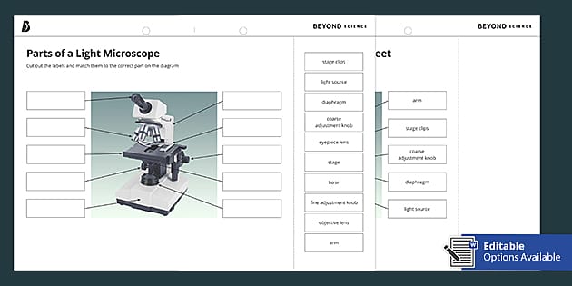
Parts of a Light Microscope Cut and Stick Worksheet - Twinkl

Compound Microscope Parts – Labeled Diagram and their ...
Microscope

Compound and Stereo- microscopes - Microscopes 4 Schools
Microscope Parts & Specifications | Microscope World Resources

Omano Microscope for Students 40x to 400x Full-Size Monocular Compound Professional Student Microscope for Science, Laboratory, Classroom, Biology, ...
Microscope Fill In The Blank - Fill Online, Printable ...
Optical Microscopes – Some Basics | Science Lab | Leica ...
Transmitted light microscope B3 Professional series B3-220ASC ...

Parts of Stereo Microscope (Dissecting microscope) – labeled ...
Parts of a Compound Light Microscope
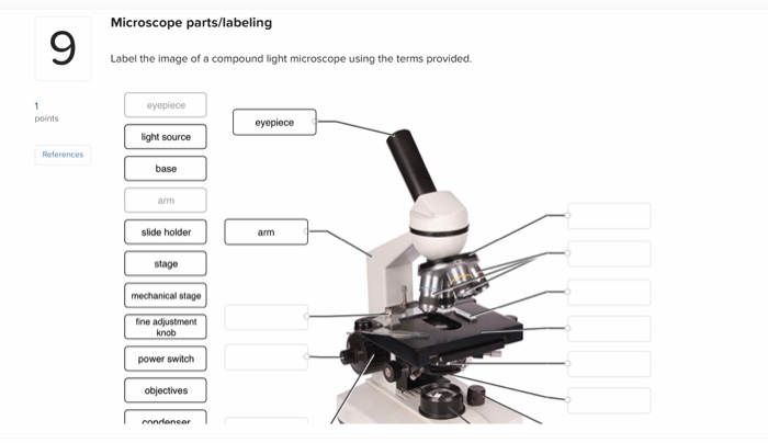
Solved Microscope parts/labeling 9 Label the image of a ...
Molecular Expressions Microscopy Primer: Specialized ...
4,897 Microscope Labeled Images, Stock Photos & Vectors ...

Microscope Parts and Functions
Post a Comment for "39 light microscope with labels"